Published: Thursday, August 31, 2023 – 12:02
New NIST Measurements Aim to Advance Portable MRI Technology
Iron oxide nanoparticles also offer the advantage that they are broken down by the human body instead of potentially accumulating in tissue, notes NIST researcher Samuel Oberdick. By comparison, a small amount of gadolinium may accumulate in tissue and could confound the interpretation of future MRI scans if it’s not taken into account.
Toward these ends, researchers measured the properties of brain tissue at low magnetic field strength. Their results were published in the journal Magnetic Resonance Materials in Physics, Biology and Medicine.
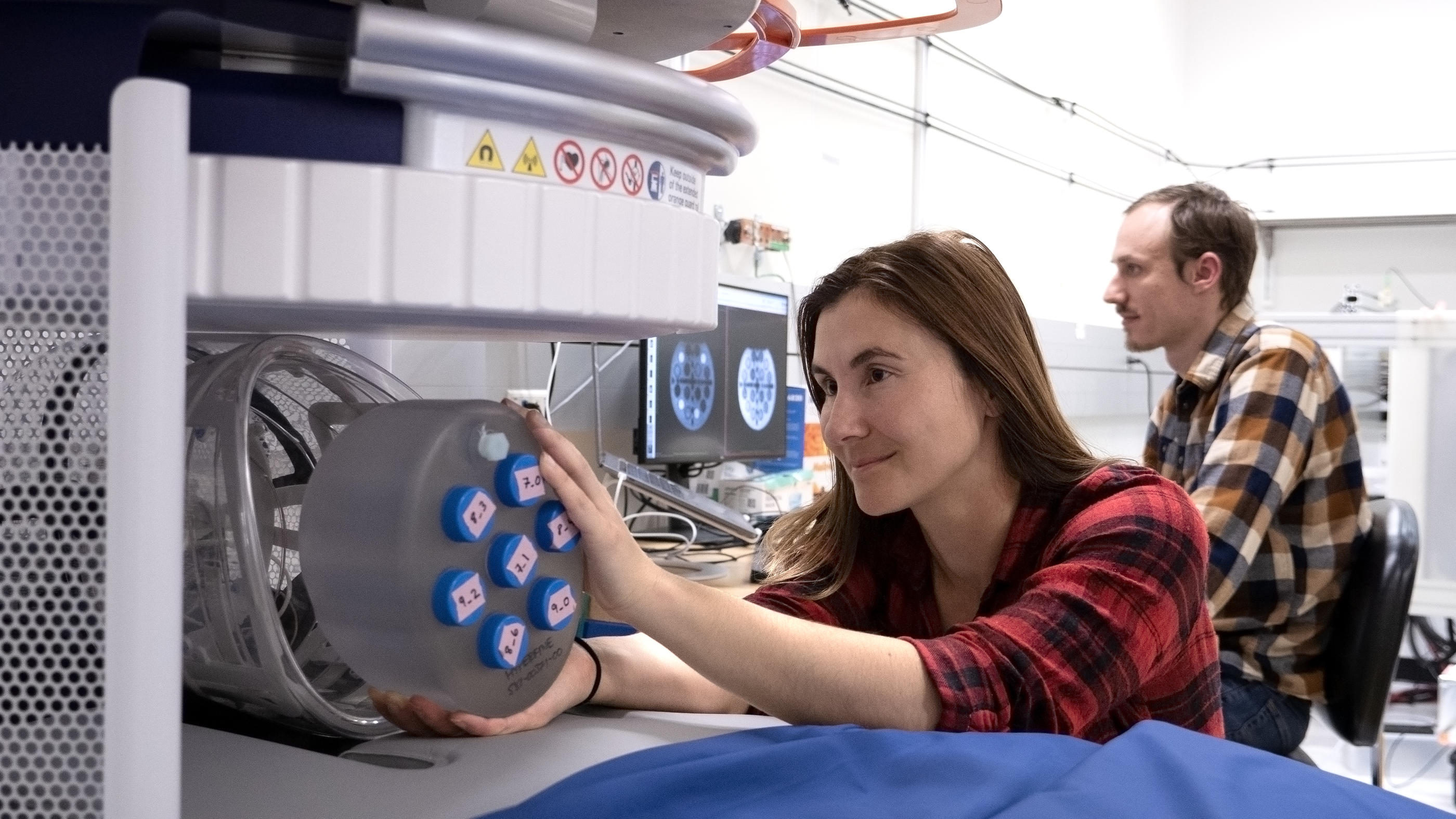
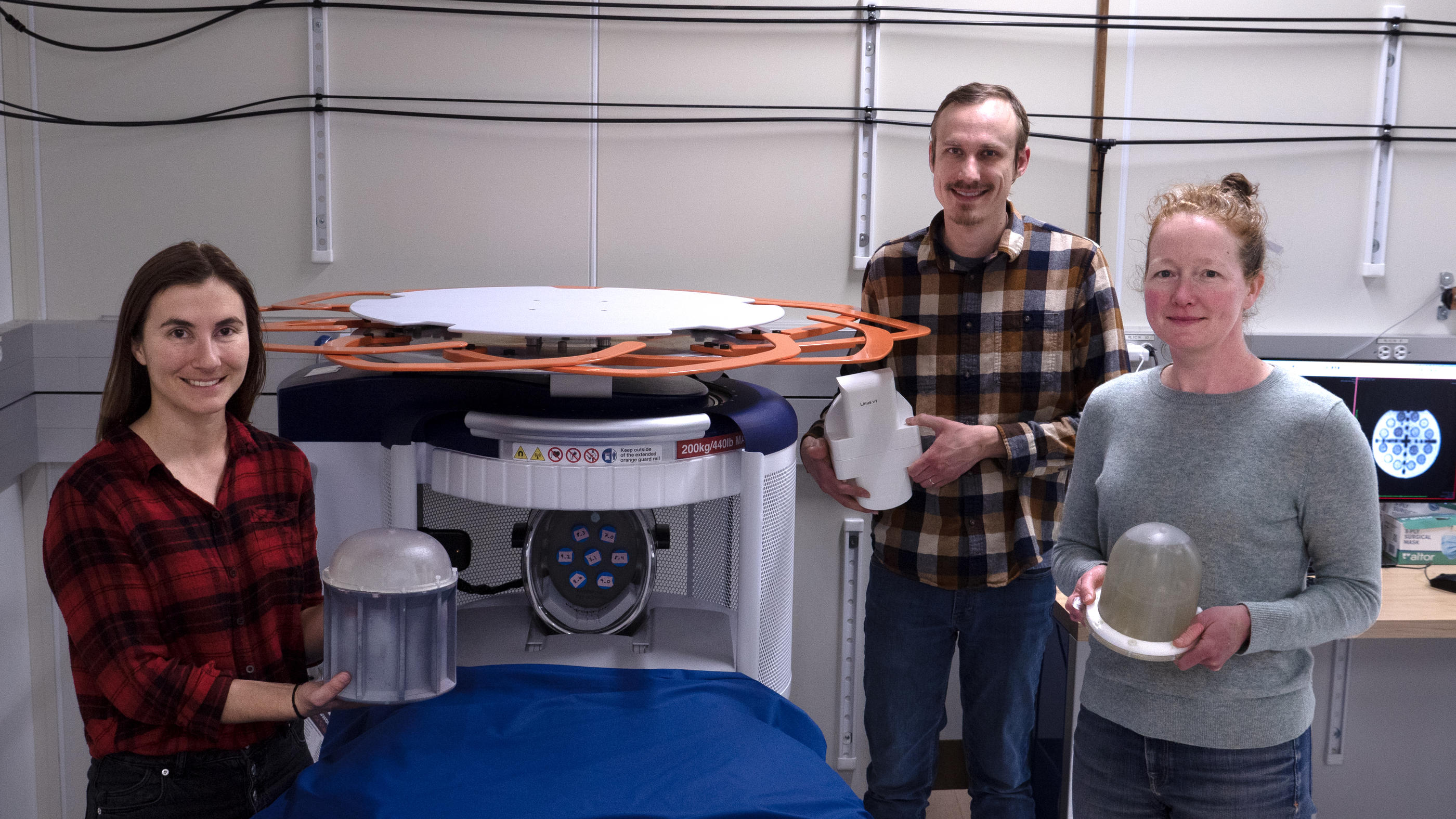
NIST researchers Kalina Jordanova and Stephen Ogier conduct MRI measurements using a magnetic field strength of 64 millitesla (mT), which is at least 20 times lower than the magnetic field in conventional MRI scanners used in hospital settings. Credit: R. Jacobson/NIST
MRI contrast agents—magnetic materials that are injected into patients and enhance image contrast—make it easier for radiologists to identify anatomical features or evidence of disease, and are routinely used in MRI at conventional magnetic field strengths. However, researchers are just starting to understand how contrast agents might be used with the new low-field MRI scanners. At the lower field strengths of these scanners, contrast agents may act differently than at higher field strengths, creating opportunities to use new types of magnetic materials for image enhancement.
In separate work, NIST researchers are exploring several candidate materials that can significantly boost image quality in low-field MRI scans.
“Magnetic resonance images of tissue differ depending on magnetic strength,” says NIST electrical engineer Kalina Jordanova. “With low-field MRI systems, the contrast of the images is different, so we need to know how human tissue looks at these lower field strengths.”
But for low-field MRI scanners to reach their full potential, more research is needed to understand the relationship between low-field images and the underlying tissue properties they represent. Researchers at the National Institute of Standards and Technology (NIST) have been working on several fronts to advance low-field MRI technology and validate methods for creating images with weaker magnetic fields.
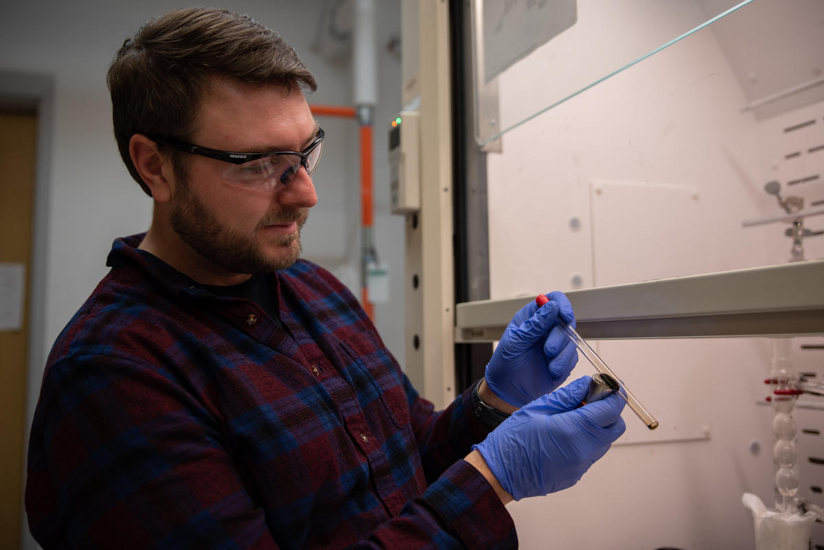
NIST scientists and their colleagues compared the sensitivity of several magnetic contrast agents in low magnetic fields. The researchers found that iron oxide nanoparticles outperformed traditional contrast agents, which are made of the element gadolinium, a rare-earth metal. At low magnetic field strength, the nanoparticles provided good contrast using a concentration of only about one-ninth that of the gadolinium particles.
However, someone has to pay for this content. And that’s where advertising comes in. Most people consider ads a nuisance, but they do serve a useful function besides allowing media companies to stay afloat. They keep you aware of new products and services relevant to your industry. All ads in Quality Digest apply directly to products and services that most of our readers need. You won’t see automobile or health supplement ads.
Magnetic resonance imaging (MRI) machines can clearly view non-bony parts of the body—soft tissue such as the brain, muscles, and ligaments—as well as detect tumors, making it possible to diagnose many diseases and other conditions. However, the powerful magnets in conventional MRI machines make them expensive and bulky, confining them mainly to hospitals and other large facilities.
As an alternative solution, companies are developing new portable versions that have lower-strength magnetic fields. These new models can potentially expand the ways in which MRI is used. For instance, low-field MRI systems could be deployed in ambulances and other mobile settings. They also could cost much less and make MRI more widely available, including in underserved communities and developing nations.
NIST researchers collaborated with the University of Florence in Italy and Hyperfine in Guilford, Connecticut, and reported their findings in the journal Scientific Reports.
Thanks,
Quality Digest